두시하태독법의 Th2 분화 조절을 통한 알레르기 비염 완화 효과
Reduction of Allergic Rhinitis by Controlling the Th2 Differentiation of Douchi Hataedock
Article information
Abstract
Objectives
This study investigated the effects of Douchi Hataedock on Th2-skewed conditions to control allergic rhinitis.
Methods
NC/Nga mice were divided into three groups: 10 mice were assigned to the control group (CTRL; no treatment), 10 mice to allergic rhinitis-induced (ARE) without treatment group, and 10 mice to the allergic rhinitis-induced (FGT) after Douchi Hataedock treatment group. The 3-week-old mice of the FGT group were given one 10 mg/kg dose of Douchi Hataedock extract and resensitized to allergic antigens at weeks four, five, and six. Allergic rhinitis was induced primarily in mice nasal cavities for five days after one week of final sensitization. The second induction used the same method one week after the first induction was completed. After one week, the nasal mucosal tissues of each group were observed. Immunohistochemical staining for IL-4, STAT6, CD40, FcεRI, substance P, MMP-9, NF-κB p65, p-IkB, and iNOS in the nasal mucosa was also performed.
Results
The FGT group had less respiratory epithelial damage and less mucin secretion in goblet cells than the ARE group and showed a 62% decrease in IL-4, 85% decrease in STAT6, 71% decrease in CD40, 69% decrease in FcɛRI, 43% decrease in substance P, 49% decrease in MMP-9, 43% decrease in NF-kB p65, 38% decrease in p-IkB, and 73% decrease in iNOS compared to the ARE group.
Conclusions
Douchi Hataedock lessens inflammation in epithelial and goblet cells and reduces inflammatory mediator secretion in a mouse allergic rhinitis model.
I. 서 론
알레르기 비염은 특정 알레르기 항원에 대한 면역반응으로 모세혈관 확장과 분비샘 과민반응이 나타나는 염증 질환이다1. 대부분 수양성 콧물, 코막힘, 재채기, 비강 내 가려움증 등을 호소하며, 이러한 증상이 하나 또는 여러 가지가 복합적으로 나타나기도 한다2,3.
국내 알레르기 질환 발병률은 10~15%로 정도이며, 최근 환경오염과 스트레스 등으로 인한 면역력 저하로 알레르기 비염을 비롯한 다양한 알레르기 질환이 증가하고 있다4. 알레르기 비염 치료는 소염제, 휴식, 수분섭취, 습도 및 온도조절 등의 대증요법이며, 만성인 경우에는 보존적 치료나 수술적 처치로 증상을 완화시킬 수 있지만 근본적인 치료법이 확립되어 있지 않다5.
알레르기 비염에 관한 국제적 치료 가이드라인 (Allergic rhinitis and its impact on asthma, ARIA)6에서는 치료법으로 회피요법, 스테로이드 주입, 항히스타민제, 약물요법, 면역요법, 아래코선반 절제술, 레이저 수술 등을 제시하였다. 이러한 치료법들은 여러 가지 부작용을 유발할 수 있다. 약물요법은 과민반응, 어지럼증, 두통, 불면증, 빈맥 등의 부작용이 생길 수 있으며, 항히스타민제는 소아에게 사용할 수 있는 근거가 부족하고, 장기간의 스테로이드제 사용은 성장장애를 유발할 수 있다6. 특정 항원을 이용하는 면역요법의 경우에는 국소부종, 두드러기, 아나필락시스를 일으킬 위험이 있다7. 이러한 치료의 한계로 대부분 증상 재발이 반복되면서 한방치료를 병행하고 있는 추세이다8. 알레르기 비염은 환자가 항원에 대한 면역력이 약하고 알레르기 소인이 있을 경우 재발률이 높고 다른 질환으로 이환되거나 합병되기 쉽다9.
소아에서의 알레르기 비염은 태아에서 발생하는 Th2 skewed condition과 연관이 있다10,11. 임신 중에는 모체 내에서 임신 상태를 유지하기 위한 면역 기전의 변화가 일어나서 Th2 cytokine이 Th1 cytokine보다 과발현하게 된다12. 이러한 변화는 알레르기 비염 같은 알레르기 질환에서 발생하는 변화와 일치하며, 출생시 모체의 불균형적인 면역체계 영향을 받아 알레르기 비염과 같은 Th2 분화 주도 염증성 질환이 발병하게 된다13.
하태독법은 두시법(豆豉法), 감초법(甘草法), 황련법(黃連法), 주밀법(朱蜜法) 등이 있으며, 한약재를 달인 약물을 부드러운 천이나 비단에 묻혀 입안의 더러운 것들을 닦아주면서 소량 먹이는 방법이다14. 두시하태독법과 관련된 이전 연구14-16에서 두시하태독법을 시행한 Nc/Nga mouse에서는 아토피피부염에서 보여지는 과도한 Th2 분화가 감소하였으며 염증으로 인한 피부손상도 적었다. 이러한 결과를 보면 Th2 skewed condition 조절을 통하여 알레르기 비염에도 예방적 효과가 있을 것으로 생각된다.
이에 본 연구는 하태독법의 Th2 skewed condition 조절을 통한 알레르기 비염 완화 효과를 알아보고자 하였다.
II. 재료와 방법
1. 두시 추출물 제조
두시(duochi, fermameted Glycine max Merr.; 남영제약영농조합, Muju, Korea)는 서리태(Glycine max Merr.)를 청호(Artemisia Apiacea Herba)와 상엽(Mori Folium) 2가지 약재를 1:1로 한 전탕액에 삶은 후, 5일간 37~38 °C에서 발효시킨 다음 열풍으로 건조시켰다. 두시 열수추출물은 두시 100 g을 파쇄하여 1000 ml의 증류수에 넣고 3시간 동안 전탕하여 여과하였다. 그 여액을 rotary evaporator에서 50 ml으로 농축한 후 동결 건조하여 추출물 15 g(수득률 15%)을 얻었다.
동일 시료로 수행된 이전 연구에서의 HPLC 결과에 따르면 두시에 포함된 isoflavone은 genistin, genistein, daidzine, daidzein이다. 일반 콩에 비해 genistin, genistein, daidzein는 증가하였고, daidzine은 감소하였다14.
2. 실험동물
실험동물은 태령 3주된 Nc/Nga 수컷 생쥐(중앙실험동물, Seoul, Korea)를 사용하였다. 대조군(Ctrl), 알레르기 비염 유발군(ARE), 두시하태독법 시행후 알레르기 비염 유발군(FGT)으로 나누었다. 각 군에 10마리씩 배정하였다. FGT는 태령 3주에 두시 추출물 10 mg/kg을 1회 경구 투여하는 하태독법을 실시하였다.
3. 알레르기 비염 유발
ARE와 FGT는 하태독법 실시 4주, 5주, 6주에 각 1회씩 ovalbumin(OVA : chicken egg albumine, grade V, Sigma)을 항원으로 사용하여 0.1% OVA용액(OVA 25 μg, 수산화알루미늄겔(Al(OH)3 gel) 1 mg, 인산완충용액(phosphate buffered saline, PBS) 300 μl을 복강주사하여 감작시켰다. 최종 감작 후 1주 후 5일 동안 OVA 100 μg, PBS 20 μl을 생쥐 비강 내에 점적하여 1차로 알레르기 비염을 유발하였다. 1차 유발 완료 후 1주 후 동일한 방법으로 2차 유발하였다(Fig. 1). 유발 확인은 콧물과 코 긁기 행동을 통하여 확인하였다. 본 연구는 부산대학교 IACUC 승인을 받았으며(IACUC number: PNU-2016-1187), 실험실 동물의 관리와 사용은 NIH guideline에 따라 시행되었다.

Protocol for allergen sensitization and challenge.
Hataedock using fermameted Glycine (FG) was orally administered on week 3th. Mice were sensitized on week 4th, 5th, and 6th by intraperitoneal injection of ovalbumin (OVA) emulsified in alum hydroxide (Alum) (OVA 25 μg : Alum 1 mg : PBS 300 μl). On week 7th, the mice were challenged with OVA (OVA 100 μg : PBS 20 μl) intranasally. On week 9th, the mice were challenged with OVA (OVA 100 μg : PBS 20 μl) intranasally. Ctrl : no treatment group, ARE : allergy rhinitis elicited group, FGT : Hataedock using fermameted Glycine treated group before allergy rhinitis elicitation
4. 조직절편 제작
각 군을 펜토바비탈나트륨(sodium pentobarbital) 용액으로 마취하였다. vascular rinse와 10% 중성 포르말린용액(neutral buffered formalin, NBF)으로 심장관류고정을 실시하였다. 머리를 분리하고 비강 주변 구조물을 제거하였다. 실온에서 24시간 동안 10% NBF에 고정한 후 탈회액(decalcification solution, BBC, UK)에 12시간 처리하였다. 세척한 후 파라핀에 포매하여 5 μm 두께로 연속절편을 만들었다.
5. 조직화학
아래코선반의 호흡상피에서 중성점액질(neutral mucin)을 분비하는 점액분비세포의 변화를 조사하기 위하여 periodic acid-schiff reaction stain(PAS)를 실시하였다. Periodic acid에서 5분간 반응시킨 후 Schiff reagent에서 15분 동안 처리하였다. 이후 sulfurous rinse에서 2분씩 3회 세척한 후 Hematoxylin에서 1분 동안 대조염색하였다.
6. 면역조직화학
Proteolysis를 위해 proteinase K(20 μg/ml)에 5분 동안 처리한 후 10% normal mouse serum에서 2시간 동안에서 blocking 시켰다. 1차 항체인 goat anti-IL-4(1:200, Santa Cruz Biotechnology, Inc., Dallas, TX, USA), goat anti-CD40(1:50, Santa Cruz Biotec), goat anti-FcɛRI receptor(1:50, Santa Cruz Biotec), goat anti-substance P(1:50, Santa Cruz Biotec), goat anti-MMP-9(1:50, Santa Cruz Biotec), goat anti-NF-kB p65(1:50, Santa Cruz Biotec), goat anti-p-IκB(1:250 Santa Cruz Biotec), goat anti-iNOS(1:100, Santa Cruz Biotec)에 4 °C humidified chamber에서 72시간 동안 반응시켰다. 이후 2차 항체인 biotinylated mouse anti-goat IgG (1:100, Santa Cruz Biotec)에 실온에서 24시간 link 하였다. 그런 다음 avidin biotin complex kit (Vector Lab, USA)에 실온에서 1시간 동안 반응시켰다. 0.05M tris-HCl 완충용액(pH 7.4)에서 발색시킨 후, hematoxylin으로 대조염색하였다.
7. 영상처리
면역조직화학 결과는 영상분석을 통해 수치화 (means±standard error) 하였으며, image Pro Plus (Media cybernetics, USA)를 이용하였다. 각 군의 표본에서 임의로 선정된 코점막 10개를 x400배율에서 촬영한 다음 10,000,000 pixels를 영상분석 하였다.
8. 통계처리
영상분석 결과의 통계는 SPSS software(SPSS 23, SPSS Inc., USA)를 이루어졌으며, one-way ANOVA 시행을 통해 유의성(P<0.05)을 검증하고 Duncan’s multiple range test로 사후 검증하였다.
III. 결 과
1. 코 점막 손상
PAS 염색 결과, ARE군와 FGT군는 알레르기 비염 유발 후 아래코선반에서 호흡상피의 손상(*)이 증가하였다. 특히 분비소낭이 없는 술잔세포(arrow)가 다수 관찰되었다. FGT군은 ARE군에 비해 호흡상피의 손상이 적었으며, 술잔세포의 점액분비도 감소하였다(Fig. 2).
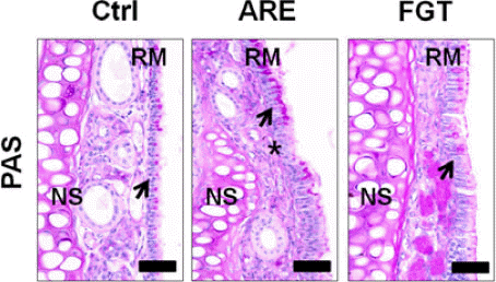
The alleviation of AR induced respiratory epithelial damages by HTD using Douchi (PAS).
Ctrl : no treatment group, ARE : allergy rhinitis elicited group, FGT : Hataedock using Douchi (fermameted Glycine) treated group before allergy rhinitis elicitation, NS : nasal septum, RM : respiratory nucosa, arrow : goblet cell, asterisk : respiratory damaged region, bar size : 50 μm
2. Th2 분화 조절
아래코선반의 점막에서 IL-4은 세포질에서 강한 양성반응을 보였다. FGT군은 ARE군에 비해 62% 유의성 있게 감소하였다. STAT6은 세포질에서 강한 양성반응을 보였다. FGT군은 ARE군에 비해 85% 유의성 있게 감소하였다. CD40는 세포질에서 약한 양성반응을 보였다. FGT군은 ARE군에 비해 71% 유의성 있게 감소하였다(Fig. 3).
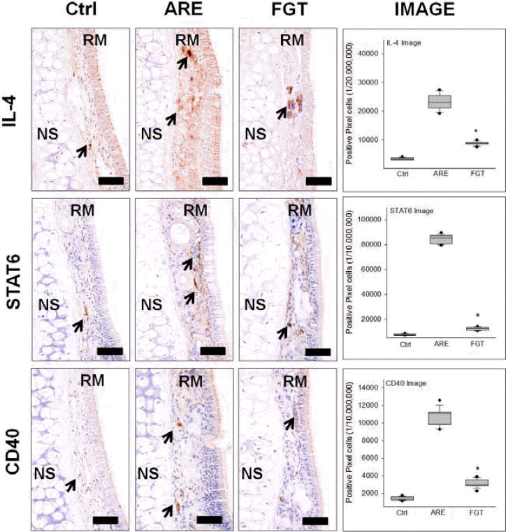
The regulation of Th2 skewed condition.
The IL-4, STAT6, and CD40 positive reaction decreased in the FGT compared with the ARE group (immunohistochemistry; bar size, 50 μm). Data of image analysis was also shown same result (*p<0.05, compared with the ARE). Ctrl : no treatment group, ARE : allergy rhinitis elicited group, FGT : Hataedock using Douchi (fermameted Glycine) treated group before allergy rhinitis elicitation, NS : nasal septum, RM : respiratory nucosa, arrow : goblet cell, asterisk : respiratory damaged region, bar size : 50 μm
3. 비만세포 활성 조절
코안아래코선반내 비만세포의 활성에 관여하는 FcɛRI 양성반응에서 FGT군은 ARE군에 비해 69% 유의성 있게 감소하였다. Itching에 관여하는 substance P 양성반응에서 FGT군은 ARE군에 비해 43% 유의성 있게 감소하였다. 코점막내 부종을 유도하는 MMP-9 양성반응에서 FGT군은 ARE군에 비해 49% 유의성 있게 감소하였다(Fig. 4).
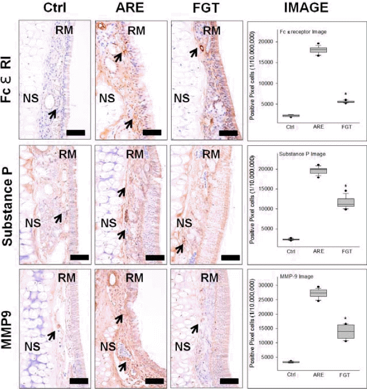
The regulation of mast cell’s activation.
The FcɛRI, substance P, and MMP-9 positive reaction decreased in the FGT compared with the ARE group (immunohistochemistry; bar size, 50 μm). Data of image analysis was also shown same result (*p<0.05, compared with the ARE). Ctrl : no treatment group, ARE : allergy rhinitis elicited group, FGT : Hataedock using Douchi (fermameted Glycine) treated group before allergy rhinitis elicitation, NS : nasal septum, RM : respiratory nucosa, arrow : goblet cell, asterisk : respiratory damaged region, bar size : 50 μm
4. 항염증효과
염증효소전사인자인 NF-kB p65 양성반응에서 FGT군은 ARE군에 비해 43% 유의성 있는 반응 감소가 나타났다. 또한 p-IkB 양성반응에서도 ARE군에 비해 FGT군에서 38% 유의성 있는 감소를 보였다. 염증효소 iNOS 양성반응에서 FGT군은 ARE군에 비해 73% 유의성 있는 감소가 나타났다(Fig. 5).
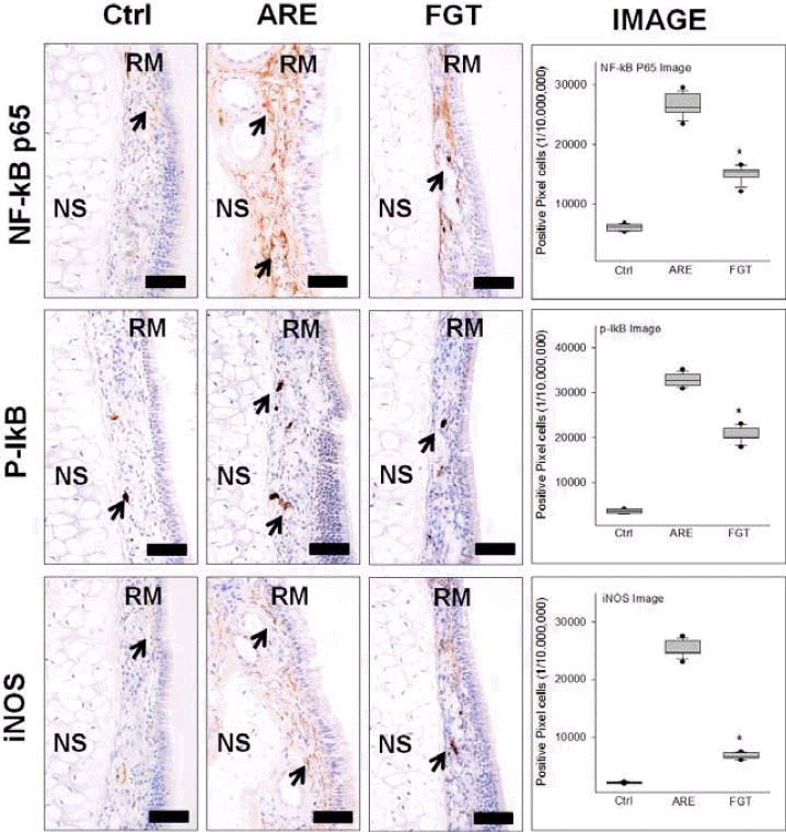
The anti-inflammation effect.
The Nf-kB p65, p-IkB, and iNOS positive reaction decreased in the FGT compared with the ARE group (immunohistochemistry; bar size, 50 μm). Data of image analysis was also shown same result (*p<0.05, compared with the ARE). Ctrl : no treatment group, ARE : allergy rhinitis elicited group, FGT : Hataedock using Douchi (fermameted Glycine) treated group before allergy rhinitis elicitation, NS : nasal septum, RM : respiratory nucosa, arrow : goblet cell, asterisk : respiratory damaged region, bar size : 50 μm
IV. 고 찰
알레르기 비염은 특정 알레르기 항원에 대한 염증 질환이다1. 최근 환경오염과 스트레스 등으로 인한 면역력 저하로 알레르기 비염이 증가하고 있다4. 최근 연구들에서는 다양한 면역학적 이상을 동반한 질환으로 보고되고 있으며17,18, 이러한 면역학적 이상은 면역계의 비정상적인 발달 과정으로 인한 Th1과 Th2 세포간의 불균형으로 Th2 반응에 치우친 과도한 면역반응으로 보고하고 있다17.
Th1 반응과 Th2 반응은 인체내에서 길항작용을 하며 균형을 유지한다. Th1과 Th2가 체내에서 어느 한쪽이 우세하게 되면 균형이 무너지고 질병이 발생할 수 있다19. Th1 유형 cytokine인 IFN-γ은 Th2의 분열을 억제하고, Th2 유형 cytokine인 IL-4는 대식세포의 기능을 억제하거나 Th1의 생성을 제한한다20,21. IL-4가 분비되면 알레르겐 항체를 만드는 B cell에서 IgE 발현이 증가된다. IL-4 분비는 Th2 분화 활성을 유도하여 CD40L 발현되며, CD40L가 B cell의 CD40과 결합함으로써 B Cell는 형질세포로 분화되어 IgE 발현이 증가된다22. 또한 Signal transducer and activator of transcription 6 (STAT6)는 전사인자로서, cytokines와 growth factors에 반응하며 IL-4, IL-13 신호에 특이적이고, 보조 T세포분화에 필수적이다23. STAT6는 Th2 분화와 IL-4의 기능에 중요한 유전자발현을 제어한다24.
본 연구에서 알레르기 비염 유발 후 두시하태독법의 비염 완화 효과를 알아보기 위해 코 점막에서의 조직 변화를 통하여 실제 확인하였다. ARE군와 FGT군는 알레르기 비염 유발 후 아래코선반에서 호흡상피의 손상이 증가하였고 분비소낭이 없는 술잔세포가 다수 관찰되었으나, FGT군은 ARE군에 비해 손상된 호흡상피가 적고, 술잔세포내 점액분비도 적었다(Fig. 2).
이러한 결과가 Th2 분화 조절과 연관이 있을 것으로 생각되어 두시하태독법에 의한 Th2 분화 조절을 알아보기 위해아래코선반의 점막에서 IL-4, STAT6, CD40의 발현 억제를 확인하였다. IL-4, STAT6, CD40은 세포질에서 강한 양성반응을 보였으며, FGT군은 ARE군에 비해 유의성 있게 감소하였다(Fig. 3). 이는 두시하태독법이 Th2 분화를 제어함으로써 알레르기 비염 유발 조절에 관여할 수 있음을 의미한다.
형질세포에 분비된 IgE는 비만세포 표면의 the surface high affinity receptors for IgE(FcεRI)와 결합함으써 비만세포의 활성화를 유도한다25,26. 활성된 탈과립형 비만세포는 histamine, prostaglandin, substance P, MMP-9, serotonin 등을 분비하여 염증 반응을 유도한다27.
Substance P는 신경 생성에 관여하는 신경펩티드(neuropeptide)28이며, 감각신경 말단에서 분비된다29. Substance P는 비만세포의 탈과립과 염증세포 증식을 유도하여 소양증을 유발한다30,31.
MMP-9은 cytokine 같은 자극에 의하여 합성, 분비되며32, laminin과 type IV collagen을 선택적으로 분해하여 기저막을 파괴하여 염증관련 세포 이주를 용이하게 한다33,34.
이에 하태독법의 알레르기 비염에 대한 완화 효과를 확인하기 위해 FcɛRI, substance P, MMP-9 의 발현 정도를 관찰하였다. FcɛRI, substance P, MMP-9 양성반응에서 모두 FGT군은 ARE군에 비해 유의성 있는 감소를 보였다(Fig. 4). 이는 두시하태독법이 비만세포에서 탈과립을 억제함으로써 알레르기 비염에서 비만세포 매개 알레르기 반응을 막을 수 있을 것으로 생각된다. 또한 substance P의 발현을 억제함으로써 소양증 완화에 효과가 있음을 의미한다.
과다한 염증세포의 이주와 침적은 알레르기 질환의 특징인데 이는 염증효소 inducible nitric oxide synthase(iNOS) 발현에 따른 nitric oxide(NO) 분비 증가의 결과이다. 산화질소(NO)는 혈관투과성의 증가를 유도하여 oxidation stress를 증가시켜 염증에 의한 조직 손상을 가속화 시킨다. 염증시iNOS의 발현은 염증효소전사인자인 nuclear factor (NF)-kB에 의해 조절되는데, 염증반응시 inhibitor of NF-kB(IkB-a)의 phosphorylation이 증가하여 염증효소전사인자 nuclear factor(NF)-kB의 핵내로의 translocation하여 들어가 target 유전자의 NF-κB binding site에 결합하여 염증관련 유전자의 발현을 유도한다35.
염증효소전사인자인 NF-kB p65와 p-IkB 양성반응에서 FGT군은 ARE군에 비해 유의성 있게 감소하였다. 또한 염증효소 iNOS 양성반응에서도 FGT군은 ARE군에 비해 유의성 있는 감소를 확인하였다(Fig. 5). 이러한 결과는 두시하태독법이 알레르기 비염에서 항염증효과가 있음을 의미한다.
하태독법의 Th2 분화 조절을 통한 알레르기 비염 완화효과를 조사하기 위해 행해진 본 연구는 태령 3주에 하태독을 시행한 Nc/Nga 생쥐에 알레르기 비염을 유발한 후 면역조직학적 변화를 관찰하였다. 하태독법 시행으로 코 점막 손상은 완화되었다. 면역조직화학적 결과, 하태독법을 실시한 군에서 IL-4, SATA6, CD40, FcɛRI, substance P, MMP-9, NF-κB p-IkB, iNOS 양성반응이 알레르기 비염 유발군보다 현저히 낮았다.
이상의 결과로 하태독법은선제적으로 Th2 skewed condition을 완화시킴으로써 염증매개물질 분비감소를 유도하여 알레르기 비염에 의한 조직 손상이 줄였다고 생각된다. 향후 후속연구를 통해 하태독법에 따른 비강점막내 Th2 condition 조절 기전 규명이 필요할 것으로 생각된다.
V. 결 론
두시하태독법의 Th2 분화 조절을 통한 알레르기 비염 완화 효과를 확인하기 위한 본 연구는 태령 3주의 약물 처리된 NC/Nga 생쥐의 아래코선반 호흡상피를 면역조직학적 변화를 통해 관찰한 결과 다음과 같은 결과를 얻었다.
두시하태독법시행군에서 알레르기 비염 유발 후 아래코선반에서 손상된 호흡상피와 술잔세포내 점액분비가 적었다
IL-4, STAT6, CD40 양성반응은 두시하태독법시행군에서 유의하게 감소하였다.
FcɛRI, substance P, MMP-9 양성반응은 두시하태독법시행군에서 유의하게 감소하였다.
NF-kB p65, p-IkB, iNOS 양성반응은 두시하태독법시행군에서 유의하게 감소하였다.
Acknowledgement
이 연구는 2016년도 정부(교육부)의 재원으로 한국연구재단의 지원을 받아 수행된 기초연구사업임(No. NRF-2016R1D1A1B03930474).